
Why Heart Failure Happens Suddenly: The “Parachute” Problem
Why does a healthy dog sometimes go into a respiratory crisis out of the blue? The rupture of the chordae tendineae...
Published on: January 18, 2026
Reviewed on: January 18, 2026
One of the most distressing experiences for a pet owner is when their loved one suddenly “crashes” into congestive heart failure. You may have known your dog had a heart murmur for years, but suddenly, their breathing becomes laboured, they are restless at night, and they are rushed to the emergency clinic. It even happens sometimes in dogs that have never had a murmur before.
Often, the culprit behind this sudden deterioration is a structural event called chordae tendineae rupture.
Most of the time, congestive heart failure due to degenerative mitral valve disease is a slow progression over many months or even years, so this very abrupt sudden type of heart failure event catches everyone out, even most cardiologists.
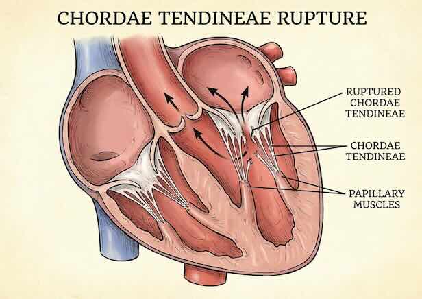
The “Parachute” of the Heart
To understand what is happening, we first need to look at the anatomy of the mitral valve. This valve acts like a one-way door between the two chambers on the left side of the heart. The left ventricle is the powerhouse of the heart, pumping blood all around the body at high pressure. So the mitral valve has to hold back this pressure, slamming shut about 120,000 times every single day. And making sure all the pumped blood goes forwards, and none leaks backwards.
To stop this door from swinging backwards when the heart contracts, it is held in place by several thin, high-strength “strings” called chordae tendineae.
Think of the mitral valve as a parachute. The valve leaflets are the fabric of the parachute, and the chordae are the suspension lines. As long as those lines are intact, the parachute stays open and catches the air. Although in this case, they are stopping the blood from leaking backwards.
Why Do the “Strings” Break?
In dogs with degenerative mitral valve disease (DMVD), the heart disease isn’t just about a “leak.” The disease actually changes the physical structure of the valve and its support strings.
There is huge natural stress on this valve tissue, and it has to constantly repair and strengthen itself to cope with the ongoing damage thatd inevitably occur. With DMVD, it seems a defective repair process is at play.
Over months or years, the collagen in these chordae is replaced by weaker, thicker tissue (myxomatous degeneration). The strings become brittle and “stretchy.” Eventually, the mechanical stress of the heart beating 120,000 times a day becomes too much, and one or more of these strings snap.
Primary vs. Secondary Rupture: What is the Difference?
The severity of the “crash” depends on which string breaks. We generally categorise them into two groups:
1. Secondary Chordae Rupture
These strings attach to the “underbelly” or the middle of the valve leaflet. Because they aren’t holding the very edge of the valve, a rupture here is often less dramatic. The valve starts to “billow” or sag backwards (prolapse), making the leak worse, but it doesn’t fail completely. This often leads to a gradual “drift” in symptoms. This is likely happening on multiple occasions over several years in a lot of dogs with DMVD, and may not be that evident when it occurs.
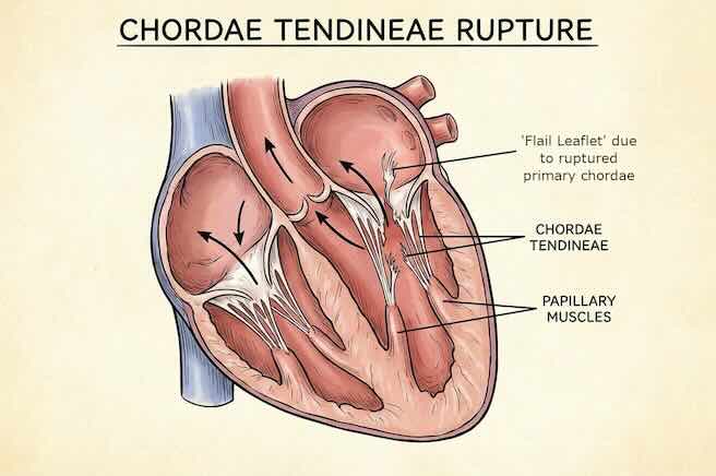
2. Primary Chordae Rupture (The “Emergency”)
These strings attach to the very edge of the valve. If a primary chorda snaps, the edge of the valve is no longer held down. It flips backwards into the left atrium every time the heart beats. This is called a flail leaflet, as it looks like a whip whizzing back and forth.
When a flail leaflet occurs, the volume of blood leaking backwards increases massively and instantaneously. The pressure in the left atrium spikes, fluid is forced back into the lungs, and the dog enters an acute state of congestive heart failure.
Why You Didn’t “Miss” the Signs
It is important to understand that a chordal rupture is a mechanical event. It is like a tyre blowout on a car. You might have known the tyre had a thin tread (the heart murmur), but the actual “pop” happens in a split second. And often the event is completely unpredictable.
This is why a dog can seem perfectly fine in the morning and be in a respiratory crisis by the afternoon.
How We Detect and Manage It
The Echocardiogram
An echocardiogram is the only way to definitively diagnose a chordal rupture. Using ultrasound, we can actually see the broken string whipping back and forth in the blood flow or observe the “flail” motion of the valve. We can also see the signs of increased left atrial pressure. This tells us that the heart’s “reserve” is gone and we need to act aggressively. For those vets getting started with echocardiography, just watch out because some of the usual markers of problems, like a large left ventrical, may not have had time to develop.
The Home Monitoring Gold Standard: SRR
While we cannot predict exactly when a string might break, we can monitor the heart’s response. If your dog has been diagnosed with DMVD, especially if they are at Stage B2 heart disease (heart enlargement but no failure yet), tracking the Sleeping Respiratory Rate (SRR) is your most powerful tool.
Check out our article on how to count the SRR, and also on monitoring devices that can help watch for these signs.
A sudden, consistent rise in the number of breaths per minute while your dog is fast asleep is often the first signal that a rupture has occurred or that the heart is no longer coping with the leak.
Emergency Treatment
Heart failure causing significant elevations is the SRR should always be seen as an emergency. Usual treatment is injectable diuretics and inodilators (pimobendan), and maybe oxgen therapy until the breathing has stabilised. Sometimes, if a particularly vital chordae has snapped, things might be unrecoverable. Only surgery would be a fix for a truly severe rupture, and no open heart centres exist currently that would be able to take an emergency case as severe as this, to my knowledge.
Summary
Chordae rupture is a natural, albeit stressful, part of the progression of advanced heart disease in dogs. By understanding that this is a structural “snapping” of the heart’s supports, we can move away from the “guilt” of missing symptoms and focus on what matters: an attempt at rapid stabilisation with diuretics (like furosemide or torasemide) and inodilators (pimobendan) to support the heart’s function. But also, acceptance that sometimes things just can’t be recovered is also important.
← Back to Blog